Medicine blended assessment

 I have been given this case to solve in an attempt to understand the topic of "Patient Clinical Data Analysis" to develop my competency in reading and comprehending clinical data including history, clinical findings, investigations and diagnosis with a treatment plan.


You can find the complete case here https://alekyatummala.blogspot.com/2020/09/45-yr-female-with-anasarca.html?m=1.


CASE:

  The presenting complaints in order of priority according to me are:

1. Pedal edema


  • Onset: insidious; on and off from 5 months back and progressive edema since 5 days.
  • Bilateral pedal edema.
  • Pitting type (Renal?Cardiac?Hepatic?Severe Malnutrition?) 
  • Progressive over a course of 1 week. Started in lower limbs and progressed to cause facial puffiness and abdominal distention. (facial puffiness hinting at renal cause? could aso be myxoedema?)
  • Aggravating factors: Walking
  • Relieving factors: on rest.
Patient went to a doctor regarding this complaint where she was  asked to reduce her fluid intake and precribed a few medicines (Name and composition unknown)

Thoughts:
Here, there is bilateral edema of pitting type which is chronic in nature as it lasted for more 72 hours. So to look at a few causes of pitting type of pedal edema:
  • Venous insufficiency (We'd require history of itching in the lower limbs,pain on walking, varicose veins)
  • Heart failure
  • Pulmonary hytertension
  • Renal disease (likely,as facial puffiness is seen)
  • Liver disease (abdominal distention is present)
  • Secondary lymphedema 
  • Anemia 
To confirm any of the above, we would require a few tests to be done:
1.Renal Function Test: Patient has high creatinine,urea and uric acid levels. This hints towards a renal pathology.
2.Liver Function Test: not done.
3.Complete Blood Picture: Microcytic hypochromic anemia is seen in the patient with Hb 6.4.

So on correlating these findings, we can suspect the cause to be a renal pathology or anemia.

2.Shortness of breath 

  • Onset: sudden and from 5 months.
  • Class: grade 3 initially but grade 4 since a week.
  • Continuous or intermittent: Continuous
Here I have a couple of more questions:
1.Was there any diurnal variation?
2.Was there any postural varaition?
3.Was it associated with cough or sputum?

On looking at the results of investiagtions, I suspect 


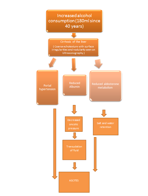
Owing to the increased Urea,Uric acid and creatinine levels, Proteinuria and hematuria leads me to think of a renal pathology which has also caused a fluid overload state resulting in pedal edema progressing to abdominal distention and facial puffiness.

Proteinuria+Edema+ Hypoalbuminemia -------> Nephrotic syndrome 
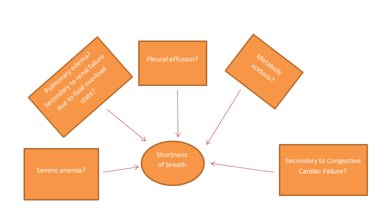
The woman has diabetes mellitus since 5 years, so here the cause of Nephrotic syndrome could be Diabetic Kidney disease or Diabetic Nephropathy.( a few causes could be ruled out because there were no amorphous deposits)So here  the reason for suspecting pleural effusion,pulmonary edema,metabolic acidosis,severe anemia and CHF and metabolic acidosis in this patient is primarily because of these constitute the common complications of Diabetic Nephropathy.


This Chest Xray shows:

  • Blunting of cardiphrenic and costophrenic angles.
Hence,Pleural effusion can be confirmed here.
Now did pleural effusion occur as a result of CHF or as a complication of ESRD?
It is more common for pleural effusion 


3.Chest pain on right side 

  • Onset: sudden since 1 week.
  • Non radiating
  • Associated with: intermittent palpitations
Thoughts:
Chest pain could be due to a cardiac,pulmonary,Gastrointestinal or musculoskeletal.
Here, I feel like cardiac and pulmonary causes are more relevant.

Since, End Stage Renal Disease due to Diabetic Nephropathy is a strong consideration here. Some of its complications or sequelae which can possibly cause chest pain are:
  • Most of the ESRD patients develop Congestive Cardiac Failure. This patient shows chest pain along with shortness of breath which can be caused by Heart Failure, bilateral pedal edema is also supportive. Congestive Heart Failure is one of the most common complications of ESRD so it is is highly suspected.
  • Pulmonary complications such Pleural effusion and Pulmoary edema can also occur due to changes in pulmonary vascular permeability and increase in pulmoary capillary pressure.Interestingly, due to heart failure also ne can develop altered pressures leading to pleural effusion or pulmonary edema.


Intermittent palpitations:
She has hypokalemia which is significant enough to cause arrhythmia and therefore palpitations. Here an ECG  would be helpful know if hyperkalemia is a culprit. 
Here the ECG shows:
  • Regular rhythm
  • Rate: 100 bpm
  • P waves present indicating sinus rhythm.


4. Past Medical History 

Patient is a known case of :
  • Diabetes Mellitus type 2 since 5 years.
  • Hypertension since 1 year.
5. On examination:

A few notable findings on examination are:
  • Pallor is present. (Suggestive of anemia)
  • Ulcer on right sole (foot ulcers are a common complication of uncontolled diabetes mellitus, so suggestive microvascular damage)
  • Blood pressure : 180/80 mm Hg 
  • Per abdomen : distended 

6. Investigations:

1.Renal Function Test:
  • Elevated urea (92mg/dl)
  • Elevated creatinine (6.4mg/dl)
  • Elevated uric acid (7.7.mg/dl)
  • Low Calcium level (8.4mg/dl)
  • High Phosphorus(6.6mg/dl)
  • Low sodium (130mEq/L)
  • Low potassium (2.9mEq/L)
Elevated urea,creatinine and uric acid indicated severe renal damage. 
Low calcium and high Phosphate are also reflections of renal damage as there is decreased conversion of 25(OH)D to 1,25(OH)2D which leads to decresed calcium aborption in the gut)

2. Complete Blood Picture
  • Microcytic hypochromic anemia evidenced on smear.

3. Complete Urine Examination
  • Presence of Albumin +4 indicated. (This reflects large amount of protein loss, and is a marker of Renal damage)
  • Presence of Pus cells
  • Presence of Epithelial cells
  • Presence of Red Blood Cells (this indicates glomerular bleeding)
4. Arterial Blood Gas Analysis
  • Blood pH is 7.19 (acidic)
  • Partial pressure of CO2 is low (indicating compensated metabolic acidosis)
  • Partial pressure of O2 is elevated. (indicating hyperventilation)
  • Bicarbonate levels are low (indicating metabolic acidosis)

On Day 2 of her admission, after treatment with oral Antihypertensives, hypoglycemic agents, Potassium supplements and injection of Sodium Bicarbonate,

She developed:
  • Pain abdomen and no urine was passed.
  • Respiratory rate has increased from 13 to 20cpm. (Maybe for compensation of metabolic acidosis but HCo2 injections were given)
  • Serum Albumin is found to be low (indcating Hypoalbuminemia)

Arterial Blood Gas Analysis:

  • Blood pH is now 7.22 (still acidic but improved)
  • Partial pressure of Co2 has increased to 22 but is still low. 
Medications given: 
  • 1.inj.HAI  according to sliding scale

    2.t.OROFER xt bd

    3.t.PAN 40 mg od

    4.inj.LASIX 40 mg iv bd if systolic bp >110mmhg

On Day 3,

The patient developed:
  • Shortness of Breath of sudden onset
  • Raised Bp 170/90 mm Hg
  • Metabolic acidosis
  • Refractory anuria
I believe the reason for sudden development of these symptoms is :
  • Worsening of fluid overload. But furosemide was given? Furosemide is a loop diuretic and it relieves fluid overload by causing diuresis but this woman has anuria so I think it worsened her situation.
"Renal hypoperfusion causes AKI, which may present itself with oliguria or anuria. Unfortunately, loop diuretics are accepted as ‘urine producers’ among physicians in ICUs and are often used. Therefore, under such conditions, the unnecessary use of loop diuretics may worsen renal blood flow contributing to AKI and worsen the clinical prognosis." (1)
  • Lower doses of Diuretics are not effective to treat fluid overload due to nephrotic syndrome.
"In nephrotics, edema is often refractory to diuretics because of low plasma protein, depletion of the intravascular compartment, decrease in the protein-bound fraction of the diuretic in peritubular blood, and increase in tubular fluid. Thus, higher doses are needed." (2)

  •  Short duration of Furosemide maybe a cause.

"Diuretic resistance may be caused by decreased renal function and reduced and delayed peak concentrations of loop diuretics in the tubular fluid, but it can also be observed in the absence of these pharmacokinetic abnormalities. When the effect of a short acting diuretic has worn off, postdiuretic salt retention will occur during the rest of the day."(3)


 --> Opthalmology referral showed moderate Hypertensive Retinopathy. (indicates uncontrolled hytertension)


On Day 4,

Arterial Blood Gas Analysis:

  • The pateint shows improvement in blood pH to 7.35.
  • The Partial pressure of CO2 has also improved.

The 2-D ECHO done shows Mitral valve regurgitation.

Questions:


 1) What is your complete anatomic and etiologic diagnosis from the data available in the patient's online record linked above? (ignore the provisional diagnosis on admission mentioned in the case report)

Ans. Anatomical diagnosis: Acute Kidney injury possibilty renal with tubular or glomerular affection.
       
Etiological diagnosis: Diabetic nephropathy with Iron deficiency anemia and Hypertensive retionopathy   

Other possible etiologies:

  • Malignant hypertension causing Nephropathy
  • As a part of cardiorenal syndrome?

The sequence of events according to me is:


2) What are the reasons for her:


Azotemia : 

  • This is biochemical abnormality in whcih there is accumulation of nitrogenous waste and creatinine in the blood.This is due to impairment of renal function of excretion of these waste products. In this case, there is oliguria due to damage to glomeruli and there is decrease in Glomerular filtration rate which leads to accumulation of creatinine and nitrogenous waste.
  • Impairment of capillary circulation in the kidney due to edema and tubular obstruction from protein casts are also possible reasons for azotemia in nephrotic syndrome pateints.


Anemia:

Anemia in this patient is due to :

  • Deficiency of erythropoietin.
  • Reduced red cell survival.
  • Reduced intake,absorption and utilization of dietary iron.
  • Blood loss in the form of haematuria.


Hypoalbuminemia:
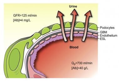
Schematic drawing of the glomerular barrier.Source: https://emedicine.medscape.com/article/244631-overview#a3
Source: https://emedicine.medscape.com/article/244631-overview#a3


Acidosis :

  • Acid base balance is maintained by excretion of acid by excretion of H+ ions and ammonium.In ESRD, there is decline in the number of functional nephrons which leads to retention of H+ ions, Impaired emmonia excretion and decreased tubular reabsoprtion of bicarbonate.
  • Insulin deficiency also leads to acidosis as when the body is unable to utilize glucose as energy source, it uses amino acids and triglycerides.Due to lipolysis, there is a rise in level of glycerol and free fatty acids. 
  • Glucagon is also released due to iniabilty to utilize glucose, which stimulates conversion of free fatty acids to ketones. The major ketoacids produced, acetoacetic acid and beta-hydroxybutyric acid, are strong organic acids that create metabolic acidosis.


3) What was the rationale for her treatment plan detailed day wise in the record? 

Particularly mention rationale and  efficacy for some of the drugs administered such as oral and iv bicarbonate? When is iv or oral bicarbonate indicated and why is it contraindicated in certain situations? 

Ans. 
Day 1:

1.inj.NaHCO3 100 meq/iv/stat in 100 ml NS : To treat metabolic acidosis. The main therapeutic effect of sodium bicarbonate administration is in increasing plasma bicarbonate levels, which are known to buffer excess hydrogen ion concentration, thereby raising solution pH to combat clinical manifestations of acidosis.

  • Efficacy of Sodium Bicarbonate in treating metabolic acidosis in ESRD:
"Patients supplemented with oral sodium bicarbonate are less likely to experience rapid progression of kidney disease."(4)

 "The relative risk for all-cause mortality in the second trial was 0.83 (95 % confidence interval, 0.68, 1.02) and renal replacement therapy was used less frequently in the bicarbonate group. These two broadly positive efficacy findings of the trial, however, are negated somewhat by the finding that bicarbonate therapy was associated with increased risk of hypocalcemia and hypokalemia requiring treatment (respective relative risks, 1.65 and 1.14). Data relating to any efficacious or deleterious effect on hemodynamic parameters was lacking. Overall statistical analysis revealed that certainty of the clinical efficacy of sodium bicarbonate was low." (5)


Indications of IV or Oral Bicarbonate:

  • Cardiac conduction delays
  • QRS prolongation (ex. tricyclic antidepressant poisoning)
  • Metabolic acidosis
  • Nebulized sodium bicarbonate is an excellent option to treat chemical injuries resulting from chlorine gas, especially within the pulmonary mucosa. 

Contraindications:

  • Due to rapid alkalotic effects, sodium bicarbonate is contraindicated in those with symptoms or laboratory values indicating  metabolic or respiratory alkalosis due to the potential for exacerbation of symptoms.
  • Due to its ability to alter acid-base balance and buffer pH, It can influence certain drug interactions such as with Antibiotics(Doxycycline, levofloxacin, minocycline, tetracyclines),NSAIDs/other related anti-inflammatory medications and Mesalamine, sulfasalazine. This is especially seen when both Sodium bicarbonate and these drugs are taken orally.
  • The administration of sodium bicarbonate can exacerbate hypernatremia.
  • Rapid or high dose administration of undiluted sodium bicarbonate may lead to decreased CSF pressure and intracranial hemorrhage.

2.syp.POTCHOLR  15 ml in one glass water tid: it contains Potassium chloride and is used to treat hypokalemia in this patient to prevent adverse effects of hypokalemia like arrhytmias and muscular spasms. Hypokalemia is a dangerous complication of ESRD which should be corrected.

3.w/h all  OHA,anti hypertensives : To keep diabetes mellitus and Hypertension under control respectively.


Day 2:

1.inj.HAI  according to sliding scale: Human Insulin (Regular) is gieven perhaps to gain better control of her uncontrollled diabetes, sliding scale means to adjust the dose of insuline according to pre meal glucose level.

2.t.OROFER xt bd : contains ferrous ascorbate and folic acid. Used to treat her Iron deficiency anemia.

3.t.PAN 40 mg od: 

It is well known that PPIs indirectly elevate serum gastrin levels via a negative feedback effect. Interestingly, in type 2 diabetes mellitus (T2DM) animal models, it has been reported that PPIs improved glycemic control, probably via possible effects on augmenting both serum levels of gastrin and Ξ² cell mass. (6)

4.inj.LASIX 40 mg iv bd if systolic bp >110mmhg : diuretic furosemide is a short acting loop diueretic given to alleviate  fluid overload.


Day 3:

1.inj.lasix 40mg iv bd

2.tab.dytor 20mg of po: now along with Furosemide, torsemide is also added as in ESRD patients, higher doses of diuretics are required to reduce fluid overload.

3.inj.HAI S/C according to sliding scale

4.tab.telma 40 mg od po->tab.nicardia 10 mg po/sos 

Stopped telma : Angiotensin Receptor Blocker was stopped and switched with calcium channel blocker.

"Taking the RENAAL and IDNT trials together, the suggestion is that it is safe and effective to coadminister CCBs along with ARBs in the management of hypertensive type 2 diabetics with nephropathy.

Adding a CCB to an ACE inhibitor or an ARB helps in reaching goal BP, while preserving renal function in both diabetics and nondiabetics with proteinuria." (7)


5.tab.orofer ct bd po

6.inj.erythropoietin s/c twice weekly: due to inadequate production of erythropoeitin in this patient, these injections ahve been prescribed.

7.tab.nodosis 500 mg bd po: Sodium bicarbonate tablets to perhaps counteract the metabolic acidosis.

8.tab.shelcal ct po/op: to counteract the hypocalcemia in this patient secondary to ESRD.

9.syp.potcholr 15ml in one glass water tid

Day 4

Blood is transfused and an imrovement in Hb is seen to 8.4 mg/dl.

Day 5

1.Syp.lactulose 30ml bd : this laxative is given to relieve constipation in this patient.

2.protein x powder 2tbsp in one glass milk bd: to compensate for the lost protein via proteinuria.

3.inj.MONOCEF 1gm IV bd: antibiotic


4) What was the indication for dialysing her and what was the crucial factor that led to the decision to dialyze her on the third day of admission? 

Ans. The indication to dialyse her was Overload with fluid refractory to diuresis.

The patient developed:

  • Shortness of Breath of sudden onset ( The overload with fluid resistant to diueresis could cause pulmonary edema)
  • Raised Bp 170/90 mm Hg
  • Metabolic acidosis
  • Refractory anuria
I believe the reason for sudden development of these symptoms is :
  • Worsening of fluid overload. But furosemide was given? Furosemide is a loop diuretic and it relieves fluid overload by causing diuresis but this woman has anuria so I think it worsened her situation. 
"Renal hypoperfusion causes AKI, which may present itself with oliguria or anuria. Unfortunately, loop diuretics are accepted as ‘urine producers’ among physicians in ICUs and are often used. Therefore, under such conditions, the unnecessary use of loop diuretics may worsen renal blood flow contributing to AKI and worsen the clinical prognosis." (1)
  • Lower doses of Diuretics are not effective to treat fluid overload due to nephrotic syndrome.
"In nephrotics, edema is often refractory to diuretics because of low plasma protein, depletion of the intravascular compartment, decrease in the protein-bound fraction of the diuretic in peritubular blood, and increase in tubular fluid. Thus, higher doses are needed." (2)

  •  Short duration of action of Furosemide maybe a cause.

"Diuretic resistance may be caused by decreased renal function and reduced and delayed peak concentrations of loop diuretics in the tubular fluid, but it can also be observed in the absence of these pharmacokinetic abnormalities. When the effect of a short acting diuretic has worn off, postdiuretic salt retention will occur during the rest of the day."(3)


The fluid overload refractory to diuretics and persistent hypertension inspite of antihypertensives are crucial factors in deciding emergency dialysis.


5) What are the other factors other than diabetes and hypertension that led to her current condition? 


  1. I think her Severe Anemia (Hb 6.4) played a role in her edema, shortness of breath and palpitations.
  2. Abdominal distention could also have lead to difficulty in breathing due to increased pressure on diaphragm and inhibiting it's efficient contraction. It is not a Huge factor but might have contributed.
  3. Hyperkalemia also contributes to breathing diffculties and palpitations.
  4. Malnutrition due to loss of proteins through urine can also contribute to her edematous state.
  5. Her Age,not that significant though.
"In T2DM patients, the prevalence of both eGFR and Albuminuria increase with age. "(8)
    6. Chronic Heart failure and Mitral Regurgitation developed in this patient also could cause palpitations, shortness of breath and edema.


6) What are the expected outcomes in this patient? Compare the outcomes of similar patients globally and share your summary with reference links. 


Ans. Outcomes: 
  • The patient could survive upto 3 years.
"The median survival of dialysis patients with baseline HF has been estimated to be 36 months, in contrast with 62 months for those without baseline HF . Over 80% of ESRD patients who are recently diagnosed with HF are expected to die within only three years from the time of this diagnosis."(9)
  • 5 year survival is possible too.
"The 5-yr survival in CHF was even worse: 12.5%In the study that compared different subgroups, the patients with diabetes and the elderly had worse outcome. " (10)

For better outcomes:
  • Better Hyperglycemia control could lead to better outcomes:
  • " Although neither the Diabetes Control and Complications Trial (DCCT) in type 1 diabetes nor the UKPDS in type 2 diabetes showed a reduction in cardiovascular events with intensive glycemic control, a prospective, observational component of UKPDS revealed a continuous relationship between glycemic exposure and the development of HF with no threshold of risk, such that for each 1% (absolute) reduction in glycosylated hemoglobin (HbA1c), there was an associated 16% decrease in hospitalization for HF ( Similar findings also were reported recently in a large cohort study from the US ." (11)
  • Better self care such as :medication adherence, glucose monitoring, dietary modification, physical activity, weight and stress management, restricted dietary sodium and fluid intake and symptom management
  • "In a small, qualitative meta-analysis, patients with DM and HF reported lack of knowledge, skill, and efficacy in integrating multiple self-care behaviors, which led them to prioritize some over others (eg, glucose monitoring, but not daily weights)."(12)

 Newer Therapies:

  • "trials using compounds that can break the covalent AGE cross-links, with their potential to reverse established disease, seem to be the most advanced. These agents that include alagebrium chloride (ALT 711; Alteon Inc., Parsippany, NJ), a stable derivative of N-phenacylthiazolium bromide , is under investigation for a range of potential indications that include DHF and diabetic nephropathy. Indeed, the findings of a small 23-patient DHF study were published recently showing that after 16 wk, alagebrium reduced left ventricular mass along with a trend to improvement in E/E′"(12)
  • "In a recently reported pilot phase 2 clinical study, patients with type 2 diabetes and nephropathy were administered ruboxistaurin 32 mg/d along with continued ACE inhibitor and/or ARB for 12 mo, noting a significant decrease in albumin:creatinine ratio from baseline of 24% (P = 0.02) that did not change in the placebo group (−9%; P = 0.33) . Furthermore, estimated GFR fell over 1 yr in placebo-treated patients (modified Modification of Diet in Renal Disease −4.8 ± 1.8 ml/min per yr; P = 0.009) but not in the ruboxistaurin-treated group (−2.5 ± 1.9 ml/min per yr; P = 0.185)."(12)
7) How and when would you evaluate her further for cardio renal HFpEF and what are the mechanisms of HFpEF in diabetic renal failure patients?

Ans. Evaluation for cardiorenal syndrome can be done after urinary output it restored.
  • N-terminal Pro BNP levels: more than 100pg indicates heart failure.
  • ECG and Echocardiogram can be used to check the pumping function of heart.
  • Chest Xray
Mechanisms of HFpEF in diabetic renal failure pateints are:
  • Intrabdominal and central venous line pressure elevation
  • Activation of Renin Angiotensin System
  • Sympathetic overactivity
  • Oxidative injury and endothelial dysfunction 


8) What are the efficacies over placebo for the available therapeutic options being provided to her for her anemia? 


  • Injection Erythropoietin:
"low-dose weekly SC rHuEPO administration was a safe and effective method for maintaining the hematocrit value of the stable hemodiabysis patients who volunteered for this study. Weekly SC rHuEPO maintained hematocrit value as well as conventional IV therapy, " (14)

   "The baseline hemoglobin level was higher in the rHuEPO group (9.9 +/- 1.15 g/dL vs. 9.3 +/- 1.41 g/dL, p = .02) as was the pretransfusion hemoglobin level (8.0 +/- 0.5 g/dL vs. 7.5 +/- 0.8 g/dL, p = .04). At day 84, patients receiving rHuEPO received fewer red blood cell transfusions (median units per patient 0 vs. 2, p = .05), and the ratio of red blood cell transfusion rates per day alive was 0.61 with 95% confidence interval of 0.2, 1.01, indicating a 39% relative reduction in transfusion burden for the rHuEPO group compared with placebo."(15)

  • Tablet orofer which contains same components asPhosfomin
" A total of 56 patients, who were between 18-55 years of age, with hemoglobin (Hb) 6-9 g/dl and serum ferritin <15 mg/l also complying with the inclusion and exclusion criteria in the protocol were enrolled in the study after obtaining necessary approvals and informed consent. All were dispensed with Phosfomin-XT fixed-dose combination (FDC) of ferrous ascorbate (equivalent to elemental iron 100 mg) + folic acid 1.5 mg tablet once-daily after food for eight weeks. Patients were assessed at baseline and Weeks 2, 4, 6 and 8 for change in Hb level, clinical evaluation and target Hb achievement. Results: Baseline mean Hb was 08.39 ± 0.75 g/dl, which significantly increased to 9.76 ± 0.70 g/dl (16.3%) at Week 2 and further increased to 10.70 ± 0.70 g/dl (27.53%) at Week 4. At Week 6, it increased to 11.55 ± 0.67 g/dl (37.7%) and at Week 8, it increased to 12.53 ± 1.09 g/dl. Conclusions: The present study concludes that Phosfomin-XT (ferrous ascorbate, equivalent to elemental iron 100 mg + folic acid 1.5 mg) tablet should be preferred as first choice of oral iron salts to treat IDA due to positive effect on Hb value and superior tolerability." (16)
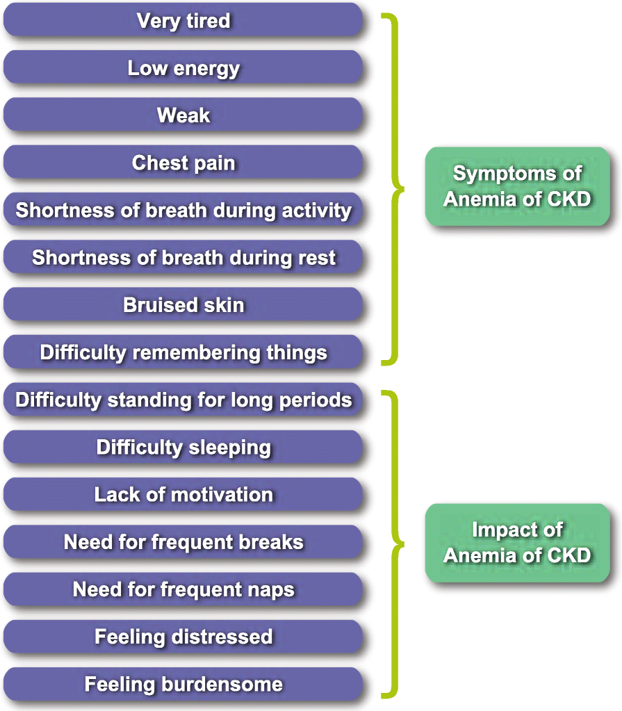
9)What is the utility of tools like the CKD-AQ that assess the frequency, severity, and impact on daily activities of symptoms of anemia of CKD? Is Telegu among the 68 languages in which it is translated? 

Ans. CKD-AQ stands for Chronic Kidney Disease-Anemia Questionnaire.
It contains 23 items covering relevant symptoms and impacts associated with anemia of CKD.

The questionnaire contains 8 items that assess the frequency of each symptom, all rated on a 5-point verbal rating scale. An additional 8 items assess the severity of these symptoms using an 11-point numerical rating scale. Five items assess the ability to do various activities, and 2 items assess the emotional impact of anemia of CKD.

Fig. 2
Source:https://jpro.springeropen.com/articles/10.1186/s41687-020-00215-8


Utility:
  • Since it is based on patient reporting, It provides relevant questions to be asked and also provides insights of the patient.
  • Some symptoms mentioned in the questionnaire like difficulty remembering things, difficulty concentrating, and restless legs were reported only after probing. This shows that providing a questionnaire makes it easy for the patient to mention all the applicable symptoms(which are mentioned in the questionnaire  after rigorously collecting data from several CKD patients.)
  • Also, this eliminates doubts of if a particular symptom is caused by CKD or another underlying cause. The patient can easily answer without having to think the symptom is irrelevant to anemia in CKD.

Languages:
    "English for Canada, UK, Australia, India, and South Africa are required. The original version of the instrument is US English." (17)


    "In parallel to Wave 2 interviews (Step 4a), a translatability assessment was conducted by experienced translators in Hindi, Russian, and Spanish to assess the feasibility of translating the draft items into other languages for use in global studies.The measure was translated into additional languages. All translations underwent either full linguistic validation, including dual forward translations and dual backward translations or linguistics review. "(18)

    10) What is the contribution of protein energy malnutrition to her severe hypoalbuminemia? What is the utility of tools such as SGA subjective global assessment in the evaluation of malnutrition in CRF patients? 

    Ans. 


    Subjective Global Assessment of malnutrition in CKD:
    quantitative scoring system consisting of 7 components with total score ranging between 7 (normal) and 35 (severely malnourished).

    Utility:

    •  In the setting of widespread chronic energy deficiency, BMI may not be a reliable marker of malnutrition. Anthropometric measurements such as skin fold thickness, mid arm circumference (MAC) and mid arm muscle circumference (MAMC) are widely used. However, the sensitivity of these methods in detecting early malnutrition, their practicability and their applicability to hemodialysis patients has not been convincing.
    • It can detect the changing nutritional status which maybe  missed by one time anthropometric and biochemical methods.
    • It is a convenient bedside tool and can be used by paramedics as well.


    2nd case 


    You can find ths case details here https://bhavyayammanuru.blogspot.com/2020/09/aki-secondary-to-uti.html?m=1


    1.Please comment on the differences in the diagnosis, therapy and outcomes in both these two patients. 

    Ans. Here the patient has a Urinary tract Infection (low grade fever, dribbling of urine,burning micturition and known case of diabetes mellitus which increases the risk).

    This patient then shows raised levels of urea,creatinine and uric acid. That means renal function is declining. So that would mean the UTI has ascended upwards and perhaps led to pyelonephritis, which is possible as there are more chances of Diabetic people developing complicated UTI.
    " A Danish study reported patients with diabetes mellitus were 3 times more likely to be hospitalized with pyelonephritis, as compared to subjects without diabetes"(19)

     Any urinary tract obstruction could also lead to acute pyelonephritis, since he is a male, there could be obstruction due to an enlarged prostrate. (Another possible  hypothesis causing acute pyelonephritis,which also seems likely as he is old)

    Decline in urinary output since 3 days+ increase in serum creatinine+pH of blood is decreased with low partial pressure of CO2===> Acute kidney injury?

    "Interstitial infiltration of neutrophils and phagocytes and extensive destruction of the parenchymal by the acute inflammatory process were found in AKI patients with acute pyelonephritis . These reports demonstrated that severe upper UTI might cause serious damage to the kidney and resulted in AKI."(20)

    Even though both the pateints have Renal failure, this patient has an early stage renal failure.Whereas, in the 45 y/o woman's case she had developed full fledged ESRD and complications as well.

    There is prolonged Aptt, thrombocytopenia and raised bilirubin which indicates some level of liver damage.


    Differences in therapy:

    nj.Piptaz 2.5mg/IV/TID---- to combat Urinary Tract Infection

    Inj.Pantop 40mgIV OD

    Inj.Lasix 40mg IV BD-- to relieve the low grade dema

    Inj.HAI S/C according to sliding scale--for glucose levels control

    Tab.Amlong 5mg OD----- Amlodipine to control the blood pressure

    Strict I/O charting

    GRBS charting 6th hrly

    Bp charting 2nd hrly

    Temp & pulse rate charting 4th hrly


    Differences in outcome:

    This patient has better prognosis than the previous patient as at this level if treated appropriately, the patient might not develop chronic kidney diease and complications.


    2.Would you agree with the provisional diagnosis shared for this  58 M in the online case report linked above?

    My hypothesis is that his UTI might have aggravated to acute pyelonephritis(suggested by shrinking of left kidney?), which has lead to Acute Kidney Injury. 

    3.What are the findings in the ultrasound of both kidneys? How do you explain those findings? Would it explain the etiology for his renal failure?

    The right kidney is normal whereas the left kidney seems smaller. Probably indicating infection in the left kidney which lead to scar formation.It does explain the etiology actually, if there is a scar due to acute pyelonephritis seen it could mean it aggravated into Acute Kidney Injury.

    Other Possible causes of unilateral small kidney:
    • Congenital small kidney
    • Reflux nephropathy due to faulty drainage mechanism
    • Renal artery stenosis
    • Glomerulonephritis (usually effects both the kidneys but rare cases)


    References:

    1)      https://www.ncbi.nlm.nih.gov/pmc/articles/PMC5656172/


    2)      https://pubmed.ncbi.nlm.nih.gov/10207256/

    3)      https://pmj.bmj.com/content/79/931/268

    4)      https://www.ncbi.nlm.nih.gov/pmc/articles/PMC4227445/#:~:text=Sodium%20bicarbonate%20infusion%20reduces%20plasma,level%20in%20serum%20%5B96%5D.

    5)      https://acutecaretesting.org/en/journal-scans/should-bicarbonate-be-used-to-treat-metabolic-acidosis

    6)      https://www.ncbi.nlm.nih.gov/pmc/articles/PMC4549663/

    7)      https://onlinelibrary.wiley.com/doi/full/10.1111/j.1524-6175.2004.4471.x#ss2-title

    8)      https://www.ncbi.nlm.nih.gov/pmc/articles/PMC5797340/

    9)      https://www.hindawi.com/journals/bmri/2014/937398/

    10)   https://cjasn.asnjournals.org/content/2/6/1186

    11)   https://cjasn.asnjournals.org/content/1/2/193

    12)   https://www.ahajournals.org/doi/10.1161/CIR.0000000000000691

    13)   https://cjasn.asnjournals.org/content/1/2/193#ref-135

    14)  https://www.researchgate.net/profile/William_Mitch2/publication/14163587_Safety_and_Efficacy_of_Low-Dose_Subcutaneous_Erythropoietin_in_Hemodialysis_Patients/links/570d3fe208ae2b772e431dbc/Safety-and-Efficacy-of-Low-Dose-Subcutaneous-Erythropoietin-in-Hemodialysis-Patients.pdf

    15)   https://pubmed.ncbi.nlm.nih.gov/16878035/

    16)   https://imsear.searo.who.int/handle/123456789/182224

    17)   https://www.valueinhealthjournal.com/article/S1098-3015(10)60784-X/pdf?_returnURL=https%3A%2F%2Flinkinghub.elsevier.com%2Fretrieve%2Fpii%2FS109830151060784X%3Fshowall%3Dtrue

    18)   https://www.ncbi.nlm.nih.gov/pmc/articles/PMC7391458/#CR17

    19)   https://www.ncbi.nlm.nih.gov/pmc/articles/PMC4346284/
                       20) https://www.ncbi.nlm.nih.gov/pmc/articles/PMC4516244/#:~:text=Interstitial%20infiltration%20of%20neutrophils%20and,kidney%20and%20resulted%20in%20AKI

    Some active learning: 
    [05/09/20, 3:51:19 PM] Srivalli: Good afternoon sir. 
    Sir I wanted to know about  the 45 y/o woman with suspected Diabetic nephropathy and nephrotic  syndrome,
    [05/09/20, 3:51:54 PM] Srivalli: 1. Was her pedal edema Bilateral? 
    2. How long did it take for the pedal edema to progress to facial puffiness? Was it within a day?
    ‎[05/09/20, 4:17:10 PM] Dr. Biswas Sir Gen Med: ‎Contact card omitted
    [05/09/20, 4:17:43 PM] Dr. Biswas Sir Gen Med: She logged the case and will be able to answer these questions better πŸ‘
    [05/09/20, 5:45:31 PM] Srivalli: Okay. Thank you sir!
    [05/09/20, 8:20:15 PM] Srivalli: Sir. This is not related to history. 
    But the patient has DM-2 since 5 years and HTN since a year, 
    So is it possible that her uncontrolled DM lead to glomerulosclerosis which lead to secondary HTN?
    [05/09/20, 8:20:30 PM] Srivalli: I mean, how would we know if it’s secondary to that?
    [05/09/20, 8:22:06 PM] Dr. Biswas Sir Gen Med: It's more likely that her diabetes and hypertension were both due to the same cause. Check out the molecular and cellular mechanisms of how visceral fat leads to hypertension and diabetes and share what you learn
    [05/09/20, 8:31:31 PM] Srivalli: Okay sir. I’ll get back to you
    [05/09/20, 9:20:19 PM] Srivalli: Sir high levels of visceral fat produce a state of chronic inflammation, here macrophages accumulate and get activated in adipose tissue and release cytokines such as TNF- alpha, MCP1 and IL-1. 
    TNF alpha disrupts the intracellular signaling from insulin receptor, sir, so this causes insulin resistance. 
    Monocyte Chemotactic Protein - it further attracts leukocytes. Increased levels of MCP cause de differentiation of adipocytes and are associated with obesity
    [05/09/20, 9:21:18 PM] Srivalli: This constant state of chronic inflammation will also cause vasoconstriction sir and this over a long period of time will translate into Hypertension.
    [05/09/20, 9:22:18 PM] Srivalli: Visceral fat is more likely to release proteins that contribute to inflammation hypertension and dyslipidemia.
    [05/09/20, 9:43:43 PM] Srivalli: Also sir, visceral fat cannot accommodate excessive lipids so it starts to store lipids in ectopic areas such as in peripheral tissues, pancreas etc where they release metabolites and exert cytotoxic effects on peripheral cells.
    [05/09/20, 9:46:21 PM] Srivalli: White adipose cells also secrete hormones with autocrine and paracrine function. Expanding fat stores leads to dynsfucntional secretion of these hormones and thereby causing metabolic impairment of insulin target tissues and also failure of insulin producing Beta cells
    [05/09/20, 10:11:12 PM] Dr. Biswas Sir Gen Med: πŸ‘πŸ‘πŸ‘
    [06/09/20, 8:27:54 AM] Srivalli: Good morning sir.
    [06/09/20, 8:32:27 AM] Srivalli: Sir in our patient with suspected diabetic nephropathy and nephrotic syndrome, 
    Her cause of SoB makes me think of 
    1.pulmonary edema( due to the fluid overload state), 
    2. Pleural effusion( chest pain with intermittent palpitations and also due to fluid overload state) 
    3. Metabolic acidosis ( due to uncontrolled DM but the Respiratory rate is normal). 

    I wanted to know how to understand whether a pleural effusion or pulmonary edema is more likely in her case?
    [06/09/20, 9:02:16 AM] Dr. Biswas Sir Gen Med: Couldn't her pleural effusion be due to the same reason as her heart failure, which is again influenced by her renal failure (aka cardiorenal syndrome) as well as the microvascular dysfunction due to her long standing diabetes (aka HFpEF)?

    One way to find out would be to get an ultrasound estimate of the volume of her pleural effusion and if tappable then go ahead with a tap to see if it's transudative in which case she could be having pleural effusion as well as pulmonary edema due to her heart failure. 

    Alekya, the intern who made her log can facilitate this. 

    Another interesting gk question around this is why do some patients of heart failure develop pulmonary edema and some develop pedal edema or pleural effusion? Hint: is it to do with the rapidity of heart failure onset? If so what are the mechanisms?
    [06/09/20, 9:24:02 AM] Dr. Biswas Sir Gen Med: Please share the online links to these quotes
    [06/09/20, 12:21:06 PM] Srivalli: Sir actually I asked the same question for last time’s case of a woman with CHF who had pleural effusion on the right side. 
    I had asked that’ the mechanisms of pleural effusion and pulmonary edema are similar but what decides what is more likely to occur? And why does pleural effusion commonly occur on right side and not left side?
    [06/09/20, 12:24:14 PM] Srivalli: Sir in pleural effusion, 
    The effusion in transudative. 
    Takes place due to change in systemic factors  such as ⬆️increase in capillary hydrostatic pressure and 
    ⬇️ decrease in colloid oncotic pressure 
    This in turn is caused due to LVF which causes increased pressure which is transmitted to the pulmonary vasculature
    [06/09/20, 12:26:33 PM] Srivalli: Similarly, for pulmonary edema to occur: 
    Due to LVF, there is increased pooling or increased pressure which causes 
    ⬆️pulmonary venous pressure 
    ⬆️ pulmonary capillary pressure 
    Hence fluid accumulates in interstitial spaces 
    ⬆️ pressure in interstitial spaces 
    Which causes accumulation of fluid in alveoli
    [06/09/20, 12:40:02 PM] Srivalli: https://www.ncbi.nlm.nih.gov/pmc/articles/PMC4800358/#:~:text=Visceral%20fat%20is%20more%20dangerous,than%20other%20indices%20of%20obesity.

    ‘Visceral fat is more dangerous than subcutaneous fat because visceral fat cells release proteins that contribute to inflammation, atherosclerosis, dyslipidemia, and hypertension.’


    https://www.ncbi.nlm.nih.gov/books/NBK279051/


    ‘“The “inflammation hypothesis” asserts that obesity represents a state of chronic inflammation where inflammatory molecules produced by infiltrating macrophages in adipose tissue exert pathological changes in insulin-sensitive tissues and Ξ²-cells.’


    ‘The “lipid overflow hypothesis” predicts that obesity may result in increased ectopic lipid stores due to the limited capacity of adipose tissue to properly store fat in obese subjects.’

    ‘The “adipokine hypothesis” refers to the principal feature of white adipose cells to function as an endocrine organ, and to secrete a variety of hormones with auto- and paracrine function.’
    ‎[06/09/20, 12:59:00 PM] Srivalli: ‎image omitted
    [06/09/20, 12:59:15 PM] Srivalli: So it’s pleural effusion sir. Can we include this in our blog?
    [06/09/20, 1:00:18 PM] Dr. Biswas Sir Gen Med: Great. Share the link to that case
    [06/09/20, 1:00:50 PM] Dr. Biswas Sir Gen Med: It's already updated in the original patient report
    [06/09/20, 1:01:54 PM] Dr. Biswas Sir Gen Med: But most of this literature sounds anecdotal than evidence based?
    [06/09/20, 1:02:29 PM] Srivalli: 35 y/o male sir 
    http://medicinedepartment.blogspot.com/2020/05/re-case-based-online-learning.html
    [06/09/20, 1:03:19 PM] Srivalli: Sir there are a few evidence based ones as well but they don’t explain the molecular mechanisms. Do I share those links too sir?
    [06/09/20, 1:03:39 PM] Dr. Biswas Sir Gen Med: The mechanism is the same isn't it? 

    How about working around the previous hint to why their expressions are different
    [06/09/20, 1:04:29 PM] Dr. Biswas Sir Gen Med: Quote the parts that you feel are evidence based with their references and we can also discuss a bit of evidence based science
    [06/09/20, 1:13:49 PM] Srivalli: https://www.ncbi.nlm.nih.gov/pmc/articles/PMC4800358/#:~:text=Visceral%20fat%20is%20more%20dangerous,than%20other%20indices%20of%20obesity 
    ‘After adjusting for age and other potential confounding factors, participants with a visceral fat mass in the upper 10th percentile had a higher odds ratio (OR) for diabetes and prediabetes than the upper 10th percentile of other adiposity indices [men, OR=15.9, 95% confidence interval (CI)=6.4–39.2; women, OR=6.9, 95% CI=3.5–13.7]. ‘
    [06/09/20, 1:15:31 PM] Srivalli: Sir there is another study in which they put the subjects on a Mediterranean diet and measure the inflammatory markers in their serum after about 16 years. 
    And it is found that those who complied with the diet, didn’t develop cognitive impairment of Type 2 diabetes.
    [06/09/20, 1:48:59 PM] Dr. Biswas Sir Gen Med: 1.the recent edema progressed within 3 days sir
    [06/09/20, 1:48:59 PM] Dr. Biswas Sir Gen Med: 2.she was not obese,wt-45kgs even 15days back..now it's 53kgs sir
    [06/09/20, 1:52:41 PM] Dr. Biswas Sir Gen Med: She's post menopause sir
    [06/09/20, 1:52:42 PM] Dr. Biswas Sir Gen Med: But then do we really know well about her trunkal obesity
    [06/09/20, 1:52:42 PM] Dr. Biswas Sir Gen Med: 6yrs back
    [06/09/20, 1:52:43 PM] Dr. Biswas Sir Gen Med: Menopause six years back was associated with AUB or was uneventful?
    [06/09/20, 1:52:43 PM] Dr. Biswas Sir Gen Med: Uneventful sir
    [06/09/20, 1:52:44 PM] Dr. Biswas Sir Gen Med: Since when is she found to have renal failure? Do we have a record of her serial RFTs before she presented to us in this admission?
    [06/09/20, 1:58:03 PM] Dr. Biswas Sir Gen Med: Pt told that she was about 60kgs before she was diagnosed with diabetes and reduced wt later on
    [06/09/20, 1:58:04 PM] Dr. Biswas Sir Gen Med: Only August reports sir
    [06/09/20, 1:58:05 PM] Dr. Biswas Sir Gen Med: They didn't have any other reports of rft  before that
    [06/09/20, 1:58:05 PM] Dr. Biswas Sir Gen Med: Yes unless we had images of her abdomen from before it would be difficult to know about her Visceral obesity
    [06/09/20, 2:28:01 PM] Srivalli: Yea sir.
    [06/09/20, 2:29:35 PM] Srivalli: So in this patient visceral fat cannot said to be the cause of the DM ?
    [06/09/20, 2:31:41 PM] Srivalli: Sir and the ulcer she has on her right sole.... can we attribute it to uncontrolled blood glucose levels?
    [06/09/20, 2:38:02 PM] Dr. Biswas Sir Gen Med: We don't know if she had the Visceral Fat. Maybe she did
    [06/09/20, 2:38:25 PM] Dr. Biswas Sir Gen Med: Does she? Is there an image
    [06/09/20, 2:40:20 PM] Srivalli: No sir but it’s written in examinatory findings
    ‎[06/09/20, 2:40:27 PM] Srivalli: ‎image omitted
    [06/09/20, 2:45:25 PM] Dr. Biswas Sir Gen Med: Let's ask for an image
    [06/09/20, 2:45:47 PM] Srivalli: Okay sir
    [06/09/20, 2:46:18 PM] Srivalli: And sir here edema is a common symptom for Heart failure, renal failure and chronic anemia. How do we differentiate what caused it?
    [06/09/20, 2:47:32 PM] Srivalli: I mean sir, why are we thinking of heart failure in this patient when her symptoms for well for ESKD itself?
    [06/09/20, 2:47:43 PM] Dr. Biswas Sir Gen Med: In all the three the common final pathway is heart failure
    [06/09/20, 2:48:50 PM] Dr. Biswas Sir Gen Med: Not shortness of breath. ESRD would produce oliguria. Again the heart failure is due to the load transferred by the kidneys
    [06/09/20, 2:50:09 PM] Dr. Biswas Sir Gen Med: Ultimately there are only two starling forces to account for edema. Hydrostatic pressure and colloidal osmotic pressure. In this lady both are jeopardized as she has nephrotic proteinuria too
    [06/09/20, 2:50:49 PM] Srivalli: Sir shortness of breath could be due to pleural effusion or pulmonary edema or metabolic acidosis?
    [06/09/20, 2:51:24 PM] Srivalli: Yeah sir. 
    And hematuria, High blood pressure, edema also point towards nephritic  syndrome no sir?
    [06/09/20, 2:52:17 PM] Dr. Biswas Sir Gen Med: Nephritic is more of acute glomerular injury while nephrotic is chronic glomerular injury. Beyond that there is not much difference
    [06/09/20, 2:53:00 PM] Dr. Biswas Sir Gen Med: Not pleural effusion as its mild just like the pericardial effusion of the other patient
    [06/09/20, 2:54:06 PM] Dr. Biswas Sir Gen Med: We really need to know when can diabetics develop acute over pre-existing chronic glomerular injury
    [06/09/20, 2:55:27 PM] Srivalli: Sir maybe due to the recently developed hypertension?
    [06/09/20, 2:55:35 PM] Srivalli: Which is out of control
    [06/09/20, 3:01:45 PM] Srivalli: Sir so if we make a timeline we can think of it like: 
    1. First she developed diabetes mellitus, which was uncontrolled. 
    2. So glycosylation of the glomerular proteins and hyalinosis lead to arteriosclerosis, and lead to END STAGE RENAL DISEASE. 
    3. It caused development and worsening of Hypertension. 
    4. Hypertension was so out of control that it caused retinopathy as well, so it could probably cause any acute episode  of hematuria?
    [06/09/20, 3:11:11 PM] Srivalli: Sir when there is diabetic nephropathy there is impairment of auto regulatory function to maintain glomerular pressure, 
    So elevated systemic pressures are transmitted to renal vasculature, which could lead to their injury and hence hematuria sir?
    [06/09/20, 3:11:24 PM] Srivalli: Sorry for bombarding with messages sir.
    [06/09/20, 4:57:55 PM] Dr. Biswas Sir Gen Med: Possible
    [06/09/20, 4:58:45 PM] Dr. Biswas Sir Gen Med: Although the sequence may not be as simple as this but yes this is a good hypothetical starting point πŸ‘
    [06/09/20, 5:06:16 PM] Srivalli: Okay sir.
    [06/09/20, 5:08:41 PM] Srivalli: Another doubt Sir, that her diabetes is causing so much havoc but her random blood sugar and HbA1c are well controlled. There is no sugar in urine sample as well. So I didn’t quite get that
    [06/09/20, 5:15:18 PM] Dr. Biswas Sir Gen Med: Once the kidneys stop functioning they cannot also excrete insulin well and so patients automatically get their sugars controlled. But would be great if you search this angle and share some evidence to support this popular hypothesis
    ‎[06/09/20, 5:16:41 PM] Dr. Biswas Sir Gen Med: ‎image omitted
    [06/09/20, 5:17:12 PM] Srivalli: Oh yeah, that makes sense sir. 
    Okay sir, I’ll look for it.
    [06/09/20, 5:17:53 PM] Srivalli: This is an indicator of uncontrolled diabetes mellitus right sir?
    [06/09/20, 5:20:26 PM] Dr. Biswas Sir Gen Med: This is an indicator of microvascular dysfunction due to diabetic microangiopathy perhaps due to adipokine induced enothelial inflammation
    [06/09/20, 5:21:27 PM] Srivalli: Okay sir. Thank you! πŸ˜€
    [06/09/20, 5:21:32 PM] Dr. Biswas Sir Gen Med: Sir... In the case... Is the renal failure primarily due to uncontrolled hypertension?
    [06/09/20, 5:21:33 PM] Dr. Biswas Sir Gen Med: Or is it a complication of diabetes sir?
    [06/09/20, 5:21:33 PM] Dr. Biswas Sir Gen Med: Like diabetic nephropathy sir
    [06/09/20, 5:21:34 PM] Dr. Biswas Sir Gen Med: Or is it correct to consider hypertension being a sequelae of renal failure sir
    [06/09/20, 5:21:35 PM] Dr. Biswas Sir Gen Med: Yes the chicken and egg conundrum that plagues most such scenarios. πŸ‘

    We expect her renal issues to be multifactorial. Her current glomerular injury manifested in nephrotic proteinuria is surely due to both diabetes and hypertension but what precipitated her current acute renal failure is still grey. Possibly she may have taken pain killers that could have precipited reduced renal perfusion
    [06/09/20, 5:23:07 PM] Srivalli: Sir I’m still somehow convinced that her acute attack is due to her hypertension as it only developed a year ago πŸ˜…
    [06/09/20, 5:33:52 PM] Dr. Biswas Sir Gen Med: πŸ‘possible but then what was the reason for her hypertension?
    [06/09/20, 5:41:21 PM] Srivalli: Sir due to diabetes lead to thickening of basement membrane of vessels so more pressure would be required to pump out the blood? The vessels get narrower. Maybe that’s how hypertension develops?
    [06/09/20, 5:43:06 PM] Srivalli: Family history of HTN is also important sir in this case. 
    And as you said, visceral fat being the common cause for the two.
    [06/09/20, 5:45:55 PM] Dr. Biswas Sir Gen Med: πŸ‘
    ‎[06/09/20, 6:52:07 PM] Srivalli: ‎image omitted
    [06/09/20, 6:55:40 PM] Dr. Biswas Sir Gen Med: πŸ‘which patient's?
    [06/09/20, 6:56:08 PM] Srivalli: Sir the 45y/o woman with suspected Diabetic nephropathy
    [06/09/20, 6:57:31 PM] Srivalli: She has hypokalemia sir, so I expected to find ST depressions πŸ™ˆ
    [06/09/20, 6:57:59 PM] Srivalli: And thought her palpitations were perhaps due to hypokalemia
    [06/09/20, 6:58:09 PM] Srivalli: But there seems to be no arrhythmia
    [06/09/20, 7:03:10 PM] Dr. Biswas Sir Gen Med: What is the sensitivity of EcG in picking up hypo or hyperkalemia?
    [06/09/20, 7:34:45 PM] Srivalli: https://www.ncbi.nlm.nih.gov/pmc/articles/PMC2390954/

    ‘Given the variability in the ECG presentation of hyperkalemia, it is not surprising that in the absence of formal criteria, the sensitivity of physician readers in the ECG diagnosis of hyperkalemia has been estimated to be as low as 0.34 to 0.43 ‘
    [06/09/20, 7:35:03 PM] Srivalli: Low sir.
    [06/09/20, 7:40:51 PM] Dr. Biswas Sir Gen Med: πŸ‘†question answered?
    [06/09/20, 7:41:18 PM] Srivalli: Yes sir. πŸ˜…
    [06/09/20, 7:41:42 PM] Srivalli: Sir but ST elevations indicate ischemia right?
    [06/09/20, 9:09:12 PM] Dr. Biswas Sir Gen Med: There are no significant ST elevations in that EcG. 
    If present they would indicate infarction rather than ischemia
    [06/09/20, 9:11:57 PM] Srivalli: Okay sir
    [06/09/20, 9:12:00 PM] Srivalli: Thank you.
    [07/09/20, 7:21:09 AM] Srivalli: ‎You deleted this message.
    [07/09/20, 7:48:10 AM] Srivalli: Good morning sir. 
    Sir this 58 year old male with AKI, is the UTI the cause for AKI? Considering it would be complicated as he is immunocpromised?
    [07/09/20, 8:26:47 AM] Dr. Biswas Sir Gen Med: Yes likely but can you detail how UTI may cause AKI
    [07/09/20, 8:28:21 AM] Srivalli: Sir since he has diabetes mellitus, it could ascend and cause acute pyelonephritis?
    [07/09/20, 8:29:00 AM] Srivalli: The interstitial infiltration of neutrophils and phagocytes can’t cause parenchyma destruction?
    [07/09/20, 8:29:54 AM] Srivalli: Sir I’m a bit confused regarding the thrombocytopenia, increased Aptt and prothrombin time and Bilirubin in this patient.
    [07/09/20, 8:32:45 AM] Dr. Biswas Sir Gen Med: What are the differentials for it?
    [07/09/20, 8:35:00 AM] Srivalli: Acute liver failure sir? But jaundice would be evident right sir?
    [07/09/20, 8:35:46 AM] Srivalli: Sir there is also an article in which it says AKI can also lead to liver damage in case sepsis but that isn’t seen here
    [07/09/20, 8:37:19 AM] Srivalli: Cholestasis sir? But the USH shows normal gall bladder
    [07/09/20, 8:57:48 AM] Srivalli: https://caseopinionsbyrollno156.blogspot.com/2020/09/medicine-blended-assessment.html
    [07/09/20, 8:59:23 AM] Srivalli: Sir my submission for the Biweekly assignment.
    [07/09/20, 9:29:39 AM] Dr. Biswas Sir Gen Med: What are the mechanisms of intrahepatic cholestasis?
    [07/09/20, 10:22:11 AM] Dr. Biswas Sir Gen Med: Please add your active learning conversations around the case in PM here to your log book as in the past. Its the active learning discussion which is more important
    [07/09/20, 2:01:43 PM] Srivalli: Sir Intrahepatic cholestatis occurs via two mechanisms mainly: 
    1. ⬆️Estrogen: it inhibits the pump of Cholic acid in hepatocytes, which transports cholic acid from hepatocyte to bile canaliculi so inspite of normal production, there is a build up of cholic acid within the cell which signals to decrease the production of bile. Conjugation of bilirubin also occurs but accumulates. 
    This increased level of estrogen is seen in cases of: usage of OCPs 
    Pregnancy 
    And intake of Anbolic steroids also has the same mechanism sir. 

    2. Neonatal hepatitis 
    There are immature mechanisms of production of bile and the liver is sensitive to injury, hence there is reduction in synthesis and flow of bile.
    [07/09/20, 2:01:49 PM] Srivalli: Yes sir

    Comments

    Popular posts from this blog

    Medicine Blended Assignment (October)

    Active learning of the topic Paraparesis